References

Dickstein K, Cohen-Solal A, Filippatos G ESC guidelines for the diagnosis and treatment of acute and chronic heart failure 2008: the Task Force for the diagnosis and treatment of acute and chronic heart failure 2008 of the European Society of Cardiology. Developed in collaboration with the Heart Failure Association of the ESC (HFA) and endorsed by the European Society of Intensive Care Medicine (ESICM). Eur J Heart Fail.. 2008; 10:(10)933-989 https://doi.org/10.1016/j.ejheart.2008.08.005

Kemp CD, Conte JV. The pathophysiology of heart failure. Cardiovasc Pathol.. 2012; 21:(5)365-371 https://doi.org/10.1016/j.carpath.2011.11.007

Kirali K, Özer T, Özgür MM. Pathophysiology in heart failure 2017. https://doi.org/10.5772/66887

Mebazaa A, Arrigo M, Parissis JT, Akiyama E. Understanding acute heart failure: pathophysiology and diagnosis. European Heart Journal Supplements.. 2016; 18:G11-G18 https://doi.org/10.1093/eurheartj/suw044

Reddi BAJ, Shanmugam N, Fletcher N. Heart failure-pathophysiology and inpatient management 2017. BJA Education. 2017; 17:(5)151-160 https://doi.org/10.1093/bjaed/mkw067

Ziaeian B, Fonarow GC. Epidemiology and aetiology of heart failure. Nat Rev Cardiol.. 2016; 13:(6)368-378 https://doi.org/10.1038/nrcardio.2016.25

Understanding the pathogenesis of heart failure

02 February 2021
Volume 32 · Issue 2

Abstract

Heart failure is a complex clinical syndrome. Jamshid Easa, Jacob Chappell and David Warriner explain the pathogenesis behind the most common form of this condition

Heart failure a complex clinical syndrome due to impairment of ventricular filling or contraction, resulting in a constellation of physical symptoms and signs, primarily due to salt and water retention. In this clinically focused review of the pathophysiology, we will summarise the main consequences of the most common type, left ventricular failure; knowledge of which is essential for those working in general practice due to the high prevalence of heart failure in the community and to aid understanding of the various pharmacotherapies that work to act on the pathological mechanisms.

Heart failure is defined as a ‘complex clinical syndrome that results from any structural or functional impairment of ventricular filling or ejection of blood’ (Dickstein et al, 2008). Patients generally present with a constellation of physical symptoms, such as fatigue and dyspnoea, and signs, such as peripheral and pulmonary oedema. It can be categorised in many ways, but the most common is left or right ventricular failure, with left ventricular failure being further subdivided into either systolic dysfunction (impaired ventricular contraction and ejection) or diastolic dysfunction (impaired ventricular relaxation and filling) (Figure 1). For the purposes of this article, we shall focus mainly on the most common, left ventricular failure (LVF), also known as left ventricular systolic dysfunction – heart failure or LVSD-HF for short (unless otherwise stated).

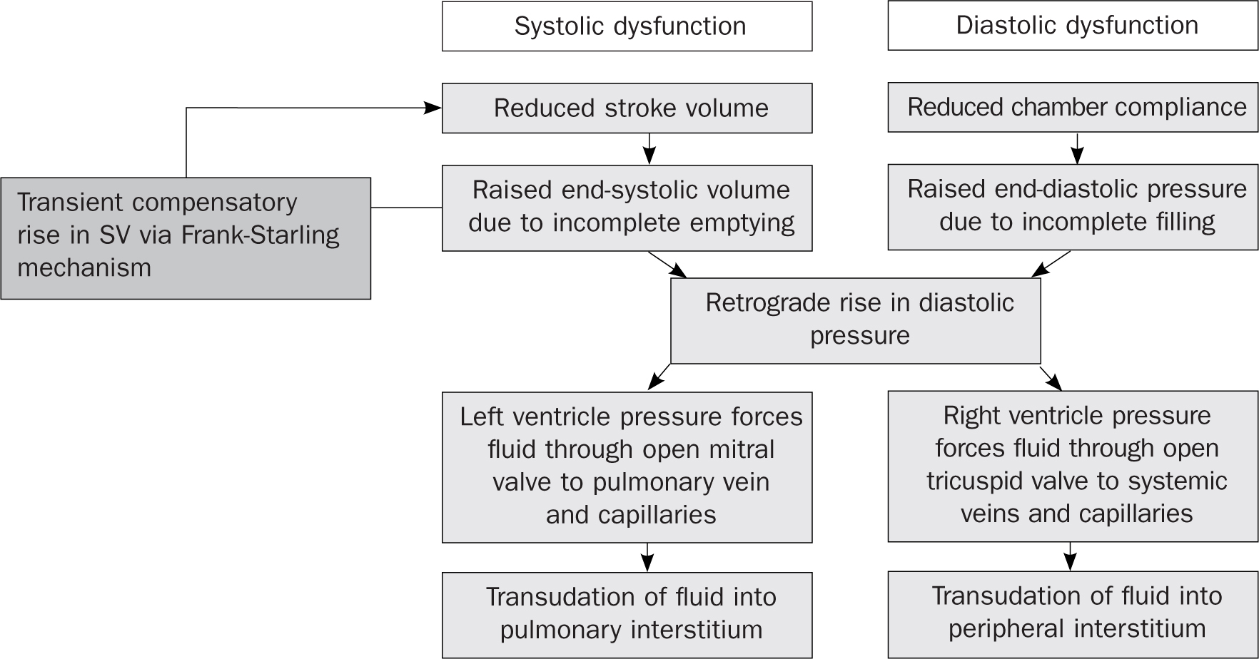
Figure 1. Mechanisms of cardiogenic pulmonary and peripheral oedema in left ventricular failure

Heart failure is a worldwide problem, affecting over 37 million individuals globally (Fonarow and Ziaeian, 2016). The lifetime risk of developing this chronic condition is 20% in adults aged 40 years and over in the developed world (Reddi et al, 2017). According to the British Heart Foundation (2020), it is estimated that as many as 920 000 people are living with heart failure in the UK and approximately around 200 000 new cases of heart failure are diagnosed every year. The prevalence of heart failure is roughly 1-2% of the adult population, which increases significantly with advancing age to more than 10% in adults over 70 years of age (Wright and Thomas, 2018).

The healthy heart

Normally, cardiac physiology is able to meet the body's metabolic demand through sufficient cardiac output (CO) defined as stroke volume (ml) eg 70 ml, x heart rate (beats per minute) e.g. 70b pm, which is typically around 5 l/min (70 x 70). Stroke volume (SV) itself is affected by three factors:

  • Preload (this is the stretch of cardiac muscle cells, also called cardiomyocytes, at the end of diastole)
  • Afterload (this is the resistance the ventricle must overcome to eject blood)
  • Contractility (this is the velocity of myocardial contraction independent of preload and afterload).

The failing heart

The reduction in CO, e.g. LVSD, is caused by loss of functional cardiac myocytes through a number of pathological conditions, with the most common being myocardial infarction, hypertension, diabetes mellitus, and ischaemic heart disease; with hypertension alone doubling the risk of developing heart failure compared to normotensive patients (Kemp and Conte, 2012). Other important but less common causes (in order of decreased prevalence) include inherited cardiomyopathies, eg dilated cardiomyopathy, infection, eg COVID-19, toxins, eg alcohol, valvular disease, eg mitral regurgitation, and arrhythmias, eg atrial fibrillation.

Failure of the heart's pumping capacity results in haemodynamic changes, which activates a succession of compensatory mechanisms in an attempt to maintain circulatory integrity by preserving central arterial pressure and thereby vital organ perfusion. While activation of compensatory measures may be beneficial in the short-term, it is the long-term sustained stimulation, as seen in chronic LVSD-HF, that leads to disease progression through ventricular remodelling and then failing to meet the demands of the body.

This leads to signs and symptoms of heart failure as a consequence of inadequate CO and inefficient venous return resulting in dyspnoea, cough and wheeze due to increased pressure in the pulmonary capillaries (Kemp and Conte, 2012). Tissue oedema, both pulmonary and peripheral, occurs when transudation of fluid from capillaries into surrounding tissues exceeds the lymphatic system's maximum drainage capacity (Mebazaa et al, 2016).

The consequence of LVSD is global hypo-perfusion of peripheral organs while causing an increase in both end-systolic and end-diastolic ventricular volumes, leading to an increase in LV end-diastolic pressure (LVEDP) and subsequently a rise in left atrial pressure, which forces fluid out of pulmonary capillaries into surrounding tissues (Kemp and Conte, 2012). The constant haemodynamic pressures cause alterations in ventricular morphology in terms of its mass, volume and composition, resulting in its overall geometry becoming less elliptical and more spherical through a process known as remodelling (Kemp and Conte, 2012). This initial hypertrophic response is a homeostatic attempt to overcome the increase in LV wall shear stress according to Laplace's law. The hallmarks of cardiac remodelling are eccentric cardiac myocyte hypertrophy and cardiac dilatation with increased interstitial matrix formation. This compensatory mechanism by the failing heart initially increases CO by increasing SV despite reduced ejection fraction (EF) but once a cut-off point is reached, it shifts to a maladaptive process, contributing to the worsening of heart failure by progressive necrosis, apoptosis and autophagocytosis of cardiomyocytes (Kirali et al, 2017). Box 1 explains EF.

Box 1.Ejection fractionIn order to understand LVSD-HF it is essential to define a measure of contractile cardiac function called the ejection fraction (EF), which is the % of blood ejected per heartbeat defined as:Ejection fraction = (end diastolic volume – end systolic volume)/end diastolic volume x 100.Confusingly, although called a fraction it is given as a % and even more confusingly normal is >55%, not 100% as you might expect. Assuming the patient has symptoms and signs of heart failure, the EF% can be used to classify patients into either reduced (<40%), eg LVSD-HF which is also known as HF with reduced ejection fraction (HFrEF), or preserved (>50%), known as diastolic heart failure or HF with preserved ejection fraction (HFpEF).

Compensatory mechanisms

The primary pathophysiological mechanism in LVSD is a reduction in SV and a subsequent fall in CO, despite a compensatory rise in heart rate. The fall in CO, in turn, leads to a decrease in mean arterial pressure (MAP) defined as cardiac output x total peripheral resistance.

The reduction in MAP causes tissue hypo-perfusion, which in turn results in activation of several compensatory measures. However, these reflexes have evolved to support the blood pressure of a healthy heart during exertion, dehydration or haemorrhage, and so they are less suited to support a failing heart (Reddi et al, 2017) and as a result the inevitable chronic up-regulation worsens the patient's clinical state in a vicious downward cycle (Conte and Kemp, 2012). Treatment of heart failure is largely trying to support cardiac function and prevent these compensatory mechanisms from running amok.

Frank-Starling mechanism

The Frank-Starling mechanism plays a key role in the early stages of LVSD-HF (Kemp and Conte, 2012) (Figure 2). The graph demonstrates three curves which represent a healthy versus a failing heart. In a healthy heart, CO increases with increasing preload, due to a greater stretch of myocardial fibres generating greater force of contraction. This is due to the unique length-tension relationship (LTR) of cardiac muscle where increasing the length of the sarcomere increases the number of possible actin–myosin interactions. Unlike skeletal muscle, cardiac muscle fibres are not at their optimum length at rest, and thus increasing venous return pre-stretches the myocardium, increasing the length of the muscle fibres, which increases the left ventricular end-diastolic volume (LVEDV), and the number of potential actin-myosin interactions and so produces a more forceful contraction, increasing the SV and CO. However, the Frank-Starling mechanism is also central to the pathophysiology of LVSD-HF, as sustained increases in venous return, combined with myocardial necrosis and scarring, cause pathological over-stretching of the myocardium, disrupting the LTR, reducing the number of potential actin-myosin interactions, and thus the force of contraction. Therefore, in a failing heart, initially SV falls due to a reduction in contractility, thus there is an increase in preload, which leads to a compensatory increase in CO. However as HF worsens, there is only a slight increase in SV for a given increase in preload (Figure 2). The curve eventually flattens and even angles downward as decompensation occurs and compensatory mechanism is exhausted with end-point being depressed CO and fluid overload.

Figure 2. The Frank-Starling Mechanism

Neuro-hormonal activation

Activation of several compensatory measures, including the sympathetic nervous system, renin-angiotensin-aldosterone system and natriuretic peptide system are aimed at optimising the heart's ailing mechanical function with subsequent improvement in oxygen delivery to peripheral tissues.

The sympathetic nervous system

The decrease in MAP is sensed by baroreceptors in the carotid sinuses and aortic arch along with mechanoreceptors in the cardiopulmonary circulation resulting in heightened sympathetic (adrenergic) activity and diminished parasympathetic (cholinergic) activity with subsequent activation of neurohormones. The sympathetic nervous system (SNS) stimulation increases release of catecholamines (adrenaline and noradrenaline), which has direct effects on the cardiovascular system by increasing HR and contractility, as well as inducing peripheral vasoconstriction, augmenting MAP by increasing total peripheral resistance (TPR). The SNS influences the myocardium through three adrenergic receptors (β1, β2 and α1) and chronic stimulation by noradrenaline induces myocyte growth and hypertrophy (Kirali et al, 2017). Commonly used in HF, β-blockers, e.g. bisoprolol, work by blocking β receptors, slowing the heart rate, lengthening the duration of diastole, improving myocardial filling and reducing myocardial oxygen demand, thereby augmentation of the cardiac pump function.

The renin–angiotensin–aldosterone system

The up-regulation of the renin–angiotensin–aldosterone system (RAAS) is another compensatory measure, which is activated through β1 and α1 receptors due to renal hypoperfusion and reduced sodium delivery to macula densa of the distal tubule. This serves to maintain intravascular volume via vasoconstriction, sodium retention and increased thirst. This is achieved by conversion of angiotensinogen to angiotensin I by renin followed by conversion of angiotensin I to angiotensin II by angiotensin-converting enzyme (ACE). Angiotensin II is a potent vasoconstrictor of systemic circulation and mediates its effects in five ways by:

  • Increasing sympathetic nervous system (SNS) activity
  • Increasing Na+ and Cl- absorption from the ascending loop of Henle
  • Stimulating the release of aldosterone from the adrenal cortex
  • Stimulating arteriolar vasoconstriction (systemic and renal)
  • Stimulating the secretion of anti-diuretic hormone from the posterior lobe of the pituitary gland.

The net effect is retention of salt and water. This increases plasma volume and hence blood pressure, which, for the short term, improves tissue perfusion. However, this short-term gain comes at a long-term cost of an increasing workload on an already failing myocardium. Figure 3 demonstrates that these neurohormonal systems also influence each other, with net result being adverse loading of the ventricles after continuous cycles of activation. Both ACE inhibitors, e.g. ramipril, and angiotensin receptor blockers (ARBs), e.g. candesartan, work to reduce the afterload on the heart by blocking this pathway, reducing total peripheral resistance and so reducing myocardial work.

Figure 3. Summarising the interplay of the body's compensatory mechanisms in heart failure

The natriuretic peptides

Natruresis means to lose salt from the body (as diuresis means to lose body water) and so the natriuretic peptides (NPs) including atrial natriuretic peptide (ANP), brain natriuretic peptide (BNP) and C-type natriuretic peptide (CNP), provide the most important counter-regulatory balance to the neurohormonal systems previously discussed by increasing sodium and water excretion. ANP and BNP are located in the atria and ventricles respectively, which are released in response to myocardial stretch due to volume expansion resulting in vasodilatation, natriuresis and diuresis (Kirali et al, 2017). CNP is mainly located in the central nervous system and acts directly on peripheral vasculature to inhibit secretion of renin, aldosterone and vasopressin. Synthetic diuretics, such as furosemide, act in conjunction with NPs to remove excess salt and fluid, which manifests as pulmonary, eg frothy cough, and peripheral oedema, eg ankle swelling. Box 2 provides more information on biomarkers.

Box 2.Blood biomarkersBecause, as mentioned, they are typically elevated in symptomatic LVSD-HF, but not asymptomatic LVSD, blood biomarkers such as BNP are sensitive for heart failure, which can be used as an aide to the diagnosis as well as determining the on-going prognosis in LVSD-HF (Table 1). Biomarkers are particularly useful in a community setting, where more advanced facilities such as echocardiography may not be readily available for use, in order to triage patients with dyspnoea of an uncertain aetiology towards appropriate one-stop shop clinics in secondary and tertiary centres.


Table 1. Natriuretic peptide levels
BNP level (pg/mol) NT-proBNP level (pg/ml) Diagnosis
<100 <400 Unlikely
100–400 400–2000 Uncertain
>400 >2000 Likely

Other changes

Like other solid organ failures, eg kidney, liver and lung failure, LVSD-HF adversely affects other systems including the endothelium, kidney, thyroid, parathyroid, bone, blood, lungs and skeletal muscle. With reference to skeletal muscle in particular, there is a loss of muscle mass, power, blood flow and mitochondrial structure and function; this sarcopenia explains why aerobic exercise, employed in cardiac rehabilitation centres, may be beneficial by seeking to reverse some of these changes.

Conclusion

LVSD-HF is a cardiac condition with a legion of causes and a myriad of effects on the body, but which ultimately all have a common purpose, serving to initially maintain organ perfusion acutely, but over time having a deleterious effect on cardiac function. The sympathetic nervous system, renin-angiotensin-aldosterone system and natriuretic peptides can all be harnessed in terms of providing targets for heart failure treatment and also to aid with diagnosis. A good working knowledge of the interplay of all these compensatory mechanisms is helpful when starting and up titrating medical therapy regardless of the clinical setting.

KEY POINTS:

  • Patients with heart failure generally present with a constellation of physical symptoms such as fatigue and dyspnoea and signs such as peripheral and pulmonary oedema
  • Myocardial infarction, hypertension, diabetes mellitus and ischaemic heart disease are all associated with heart failure
  • In most cases, the aetiology of left ventricular systolic dysfunction – heart failure (LVSD-HF) is of secondary importance to first initiating evidence-based therapies to reduced morbidity and mortality, eg ACE inhibitors and beta-blockers
  • Irrespective of the cause, the downstream pathophysiological effects and therefore the treatments, are largely the same

CPD reflective practice:

  • Which pathological conditions are associated with heart failure?
  • Can you explain why compensatory measures are beneficial in the short-term but not the long-term?
  • Why are biomarkers for heart failure particularly useful in the community?